Histology images (click to enlarge)
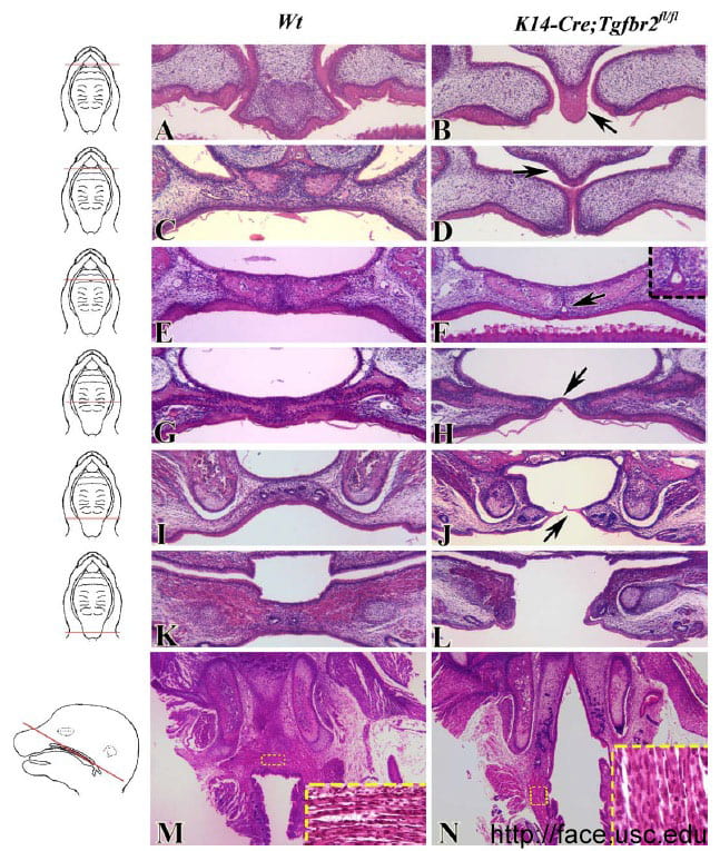
Description: Histological analyses of K14-Cre;Tgfbr2fl/fl mutant palate. (A, C, E, G, I, K) Frontal sections of the wild-type palate, from anterior to posterior. (B, D, F, H, J, L) Frontal sections of the K14-Cre;Tgfbr2fl/fl mutant palate, from anterior to posterior. (B) In the K14-Cre;Tgfbr2fl/fl sample, the primary palate epithelium overgrows to form an epithelial tongue (arrow). (D) The nasal septum fails to fuse with the secondary palatal shelves (arrow). (F) An epithelial cyst is located in the midline of the palate (arrow and insert). (H) An epithelial bridge connects the hard palate (arrow). (J) The epithelial bridge is elongated on the more posterior region (arrow). (L) On the posterior of the soft palate, there is a complete cleft. (M) Transversal section of the wild-type palate. The direction of muscle fiber is marked by a white dashed line (insert). (N) Transversal section of the K14-Cre;Tgfbr2fl/fl mutant palate. The muscle attachments of the soft palate are directed anteriorly instead of horizontally (insert).
Source: Xu et al. (2006) Cell autonomous requirement for Tgfbr2 in the disappearance of medial edge epithelium during palatal fusion. Developmental Biology 297: 238-248.
